
Let’s explore an overview of Western Blotting, a widely used technique in molecular biology and biochemistry for detecting specific proteins within a sample.
Introduction to Western Blotting: Western Blotting, also known as immunoblotting, widely used technique in molecular biology and biochemistry for detecting specific proteins within a complex mixture. This technique is valuable in understanding protein expression, post-translational modifications, and protein interactions.
It allows researchers to detect the presence and relative abundance of a particular protein of interest in a sample. It also helps in investigating changes in protein expression levels under different experimental conditions and provides insights into cellular processes and protein function.
Western Blotting finds applications in various fields of research, including cell biology, cancer research, immunology, neuroscience, and drug development. It is instrumental in studying disease biomarkers, protein-protein interactions, and the effects of different treatments on protein expression.
Materials Required for Western Blotting:
To perform a Western Blot, you’ll need several key components:
- Protein Sample: The mixture containing the proteins of interest, extracted from cells, tissues, or other biological sources.
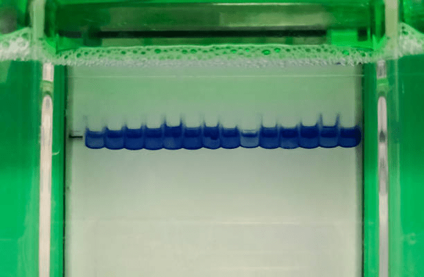
- Electrophoresis Gel: A polyacrylamide gel used to separate proteins based on their size.
- Transfer Membrane (e.g., PVDF or nitrocellulose): The membrane onto which proteins are transferred from the gel.
- Primary Antibody: A specific antibody that binds to the target protein with high affinity.
- Secondary Antibody: Conjugated to an enzyme or fluorescent tag, this antibody binds to the primary antibody, facilitating detection.
- Blocking Agent: A solution that prevents non-specific binding of antibodies to the membrane.
- Washing Buffers: To remove unbound antibodies and other unwanted components.
- Detection Reagents: Depending on the secondary antibody, these reagents enable visualization of the protein bands.
Basic Step-by-Step Western Blot Protocol:
Here’s a simplified outline of the Western Blotting process:
- Protein Separation: The protein sample is loaded onto an electrophoresis gel and subjected to an electric field, which separates the proteins based on their size.
- Transfer to Membrane: After electrophoresis, the proteins are transferred from the gel to a solid membrane, typically using a technique called electroblotting.
- Blocking: The membrane is treated with a blocking agent to prevent non-specific binding of antibodies, ensuring they only interact with the target protein.
- Primary Antibody Incubation: The membrane is incubated with the primary antibody, allowing it to bind specifically to the target protein.
- Secondary Antibody Incubation: After washing away unbound primary antibodies, the membrane is incubated with the secondary antibody, which recognizes the primary antibody and is linked to an enzyme or a fluorescent tag.
- Detection: The presence of the target protein is visualized by adding a substrate that reacts with the enzyme or a fluorescence scanner in the case of fluorescent tags.
- Data Analysis: The resulting protein bands are analyzed to determine the abundance and size of the target protein.
