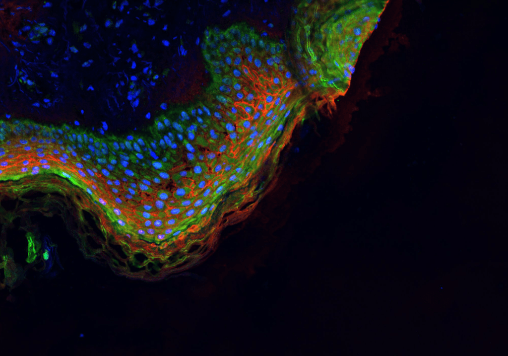
Overview
The Immunofluorescence Assay (IFA) is a widely used and versatile laboratory method that enables researchers to visualize the presence and distribution of specific antigens or proteins in biological samples. It utilizes fluorescently labeled antibodies that bind to the target molecules of interest, allowing for precise and sensitive detection. IFA provides valuable information about the spatial distribution and localization of specific antigens or proteins within cells or tissues. It is used to study protein expression patterns, cellular structures, and subcellular localization in various research areas. IFA finds extensive applications in cell biology, immunology, neuroscience, and pathology. It is instrumental in studying cellular signaling pathways, protein-protein interactions, cellular dynamics, and the effects of various treatments on cellular components.
Materials and Components in IFA:
To perform an IFA, you’ll need several key components:
- Primary Antibodies: Specific antibodies that bind to the target antigens or proteins of interest.
- Secondary Antibodies: Fluorescently labeled antibodies that recognize and bind to the primary antibodies.
- Blocking Agent: A solution used to block non-specific binding sites and reduce background noise.
- Permeabilization and Fixation Reagents: To enable antibody penetration and preserve cell/tissue structures.
- Fluorescence Microscope: The microscope equipped with appropriate filters for excitation and emission of the fluorescent labels.
Basic Steps in IFA:
Here’s a simplified outline of the IFA process:
- Sample Preparation: Cells or tissue sections are fixed and permeabilized to preserve cellular structures and allow antibody penetration.
- Blocking: Non-specific binding sites are blocked with a blocking agent to minimize background fluorescence.
- Primary Antibody Incubation: The sample is incubated with specific primary antibodies that bind to the target antigens or proteins.
- Washing: Unbound primary antibodies are washed away to reduce background signals.
- Secondary Antibody Incubation: The sample is incubated with fluorescently labeled secondary antibodies that bind to the primary antibodies.
- Washing: Unbound secondary antibodies are washed away to reduce non-specific signals.
- Mounting: The sample is mounted with a suitable mounting medium to preserve the fluorescence signal.
- Imaging: The sample is observed under a fluorescence microscope, and the fluorescence signals emitted by the bound antibodies are detected.
- Data Analysis: The images are analyzed to determine the localization and distribution of the target antigens or proteins.
Its ability to provide detailed spatial information has significantly contributed to our understanding of cellular structures and functions, advancing research in various disciplines, including cell biology, immunology, and neuroscience.